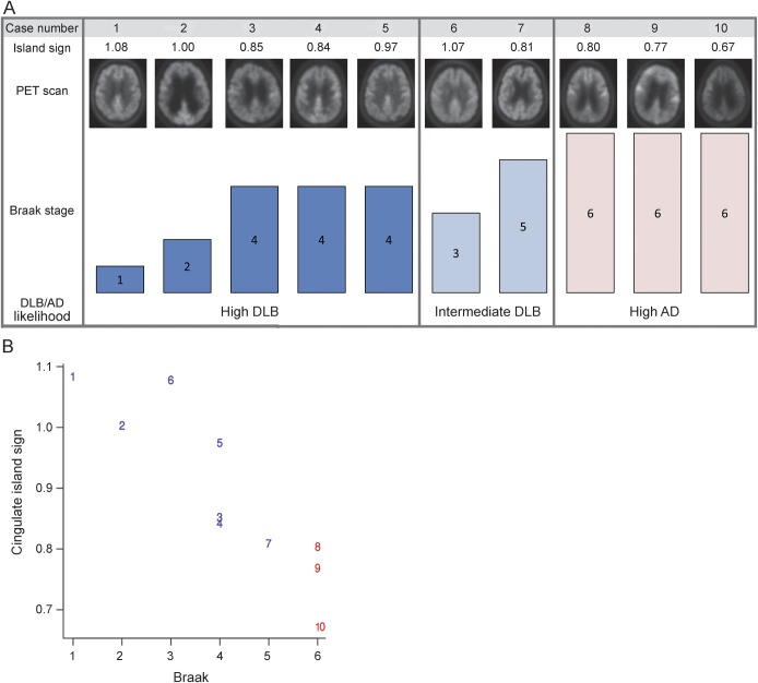
Figure 3. Pathology-confirmed clinical cases arranged by DLB/AD likelihood according to pathology and cingulate island sign.
(A) Corresponding PET scan and Braak NFT stage in rows below. Cases 1 through 6 and cases 8 and 10 had clinically diagnosed DLB, and all but cases 8 and 10 had intermediate or high likelihood of DLB at autopsy. Cases 7 and 9 were diagnosed with AD clinically. (B) Numbers represent the corresponding cases shown above; blue numbers are DLB at autopsy and red numbers are AD at autopsy. AD = Alzheimer disease; DLB = dementia with Lewy bodies; NFT = neurofibrillary tangle.

