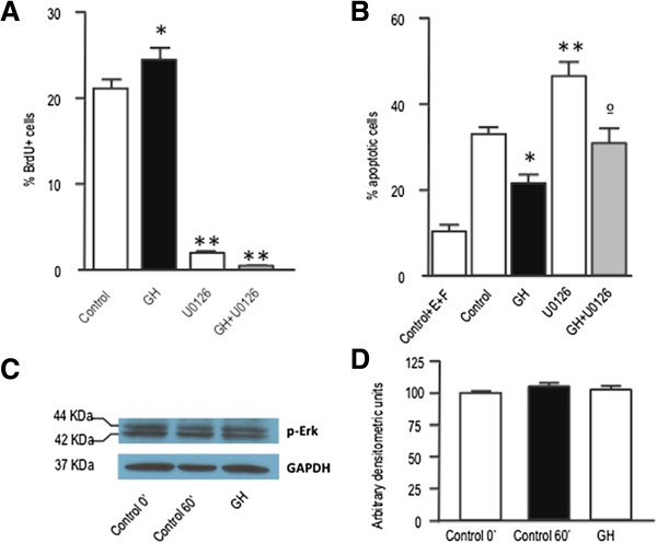
Figure 4.

ERK phosphorylation is necessary for neurosphere proliferation and survival. A) Neurospheres growing in defined media were treated for 24 h with GH (500 ng/mL), U0126 (20 μM) or GH + U0126. Control cells were treated with saline. Four hours before the end of the treatment period cells were given a BrdU pulse (10 μM). Neurospheres were then dissociated and cells were collected by centrifugation onto coated cover slips, and BrdU was detected by immunocytochemistry. * = p <0.05 vs Control; ** = p < 0.001 vs control and GH. B) Neurospheres were placed into culture plates containing defined media without EGF and FGF2, and 24 h later treated with GH, U0126 or GH + U0126, for an additional 48 h period. Control cells were treated with saline. Cell apoptosis was determined by TUNEL staining. Each bar represents the mean + SEM of 3 experiments in triplicate. * = p <0.01 vs Control; ** = p <0.001 vs control and GH; ° = p <0.01 vs GH. Basal apoptosis was determined by growing the cells in the defined media (Control + EGF + FGF2). Apoptosis values in this group were significantly lower (p <0.01) than in the other groups. E = EGF; F = FGF2. C) Neurospheres were deposited for 48 h in slides with defined media without EGF and FGF2, and then treated with GH for 1 h. Phospho ERK and GAPDH immunoreactivities were determined by western blot. D) Densitometric evaluation of results presented in C. Phospho ERK levels are expressed as arbitrary densitometric units and normalized to GAPDH levels.
