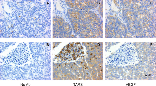
Figure 3.

TARS expression co - localized with VEGF and in leukocytes. Tissues were stained by IHC as in Figure 2. Shown are 40x images of serial sections stained with (A,B) No primary Ab as a negative control (C,D) TARS or (E,F) VEGF. Bottom panels show examples of TARS staining in infiltrating leukocytes. Bar = 50 μm. See Additional File 3 for supporting images.
