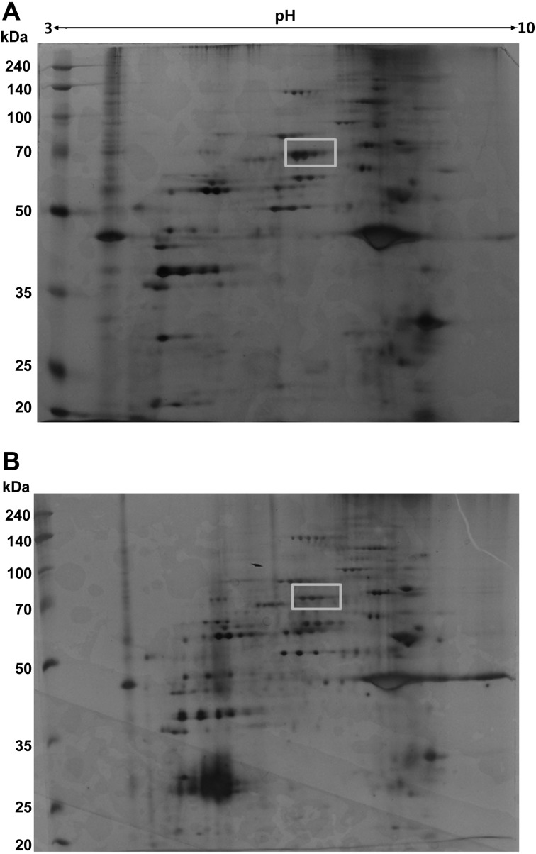
Fig. 1.
2D-PAGE analysis of PBMC extracts. Protein extracts (60 µg) from normal (A) and STZ-induced diabetic PBMC (B) were separated by IEF on Immobiline™ DryStirp pH 3–10 NL and then by SDS-PAGE. The gel was stained with silver staining. Rectangle represents the protein spot analyzed by MALDI-TOF mass spectrometry.

