Fig. 2.

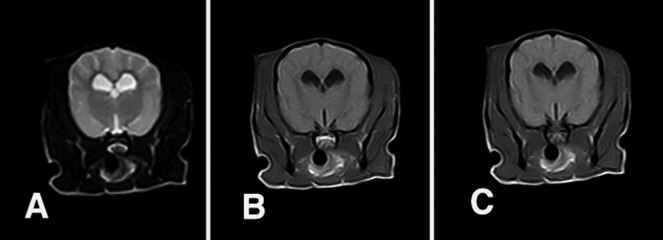
T2-weighted (A), T1-weighted (B) and T1-weighted transverse post-contrast images (C) of the brain. There were no specific findings from the MRI examination.

T2-weighted (A), T1-weighted (B) and T1-weighted transverse post-contrast images (C) of the brain. There were no specific findings from the MRI examination.