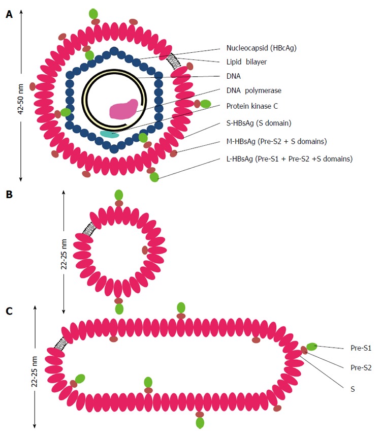
Figure 1.

Schematic representations of hepatitis B virus virion (A), spherical (B) and filamentous (C) particles. The envelope of hepatitis B virus (HBV) virion or the Dane particle (A) contains three forms of hepatitis B surface antigen (HBsAg): large (L-) HBsAg has the PreS1, PreS2 and S domains; middle (M-) HBsAg contains the PreS2 and S domains; and small (S-) HBsAg has only the S domain. The representations of the L-, M-, and S-HBsAg have no quantitative or positional significance. The L-HBsAg interacts with the viral capsid which is made of many copies of the core protein (HBcAg). The capsid encapsidates a partially double stranded DNA molecule, a DNA polymerase containing the primase and reverse transcriptase activities. The protein kinase C phosphorylates the capsid protein. The diameter of HBV virion is about 42 nm when it is stained negatively and observed under a transmission electron microscope, but it appears bigger in cryo-electron microscopy. The spherical (B) and filamentous (C) particles have a diameter about 22 nm in negative staining and appear bigger in cryo-electron microscopy. The length of the filamentous particle varies. Both the non-infectious particles contain the L-, M-, S-HBsAg and lipid.
