
Figure 1.
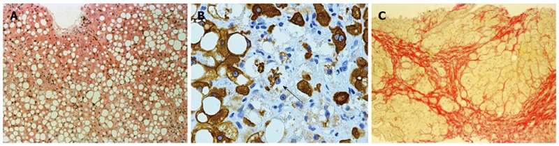
Histology of alcoholic fatty liver. A: Macrovesicular steatosis in alcoholic fatty liver (HE stain, × 218); B: Ballooned hepatocyte (arrow) containing a Mallory Denk body in alcoholic hepatitis (CAM5.2 stain for cytokeratins 8 and 18, × 218); C: Collagen surrounds nodules of hepatocytes in alcoholic cirrhosis (Serius red stain, × 872).