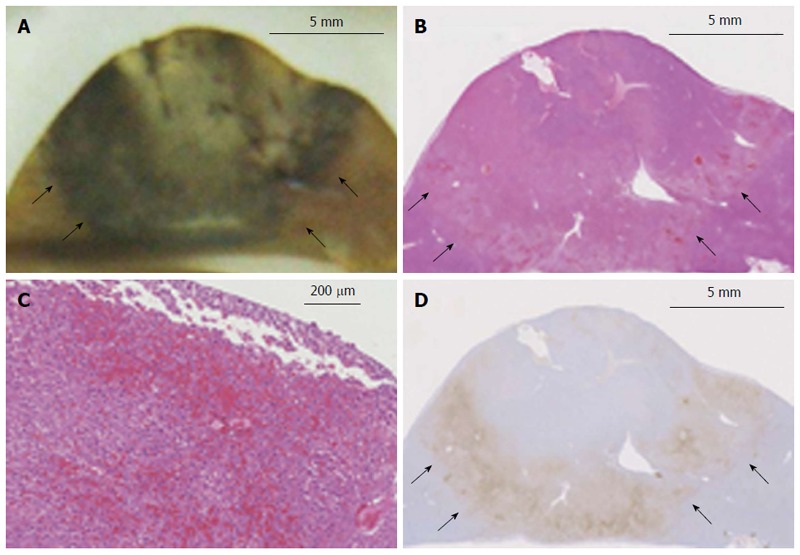
Figure 4.

Histopathological findings of a thermal lesion. A: Gross pathological examination of a radiofrequency (RF)-ablated liver shows a core area of white coagulation and a surrounding dark rim of hemorrhagic tissue (arrows). Scale bar: 5 mm; B, C: H-E staining of an RF-ablated liver shows a hemorrhagic rim surrounding the ablated area (arrows), with sinusoidal dilatation and congestion associated with cellular dissociation. Scale bar in (B): 5 mm, and in (C): 200 μm; D: Hsp70 staining of the hemorrhagic rim in (B) shows a clearly demarcated band of Hsp70 expression (arrows), which is suggestive of the presence of thermally damaged cells within the rim. Scale bar: 5 mm.
