
Figure 2. Peripheral Blood Smears in Hereditary Spherocytosis.
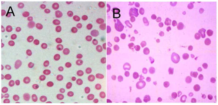
A. Typical hereditary spherocytosis. Characteristic spherocytes lacking central pallor are seen. B. Severe, recessively-inherited spherocytosis. Numerous small, dense spherocytes and bizarre erythrocyte morphology with anisocytosis and poikilocytosis associated with severe hemolysis are seen.