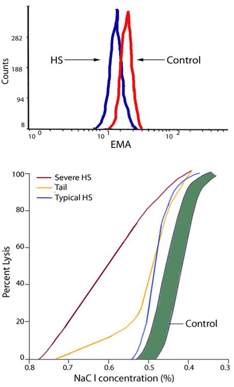
Figure 3. Testing in Hereditary Spherocytosis.

A. Eosin-5-maleimide (EMA) binding. Histogram of fluorescence of EMA-labeled erythrocytes from a normal control and a patient with typical hereditary spherocytosis. Decreased fluorescence in observed from HS erythrocytes. B. Osmotic fragility curves in hereditary spherocytosis. The shaded region is the normal range. Results representative of both typical and severe spherocytosis are shown. A tail, representing fragile erythrocytes conditioned by the spleen, is common in spherocytosis patients prior to splenectomy.
