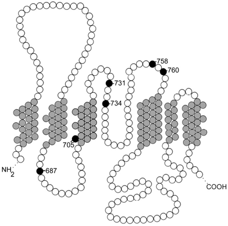
Figure 1. A partial model of band 3, the erythrocyte membrane anion transporter.

Transmembrane domains seven through twelve, shaded gray, and intracellular and extracellular loops, shaded white, are shown. Missense mutations in this region associated with ablation of anion function and abnormal cation leak, shaded black, are labeled.
