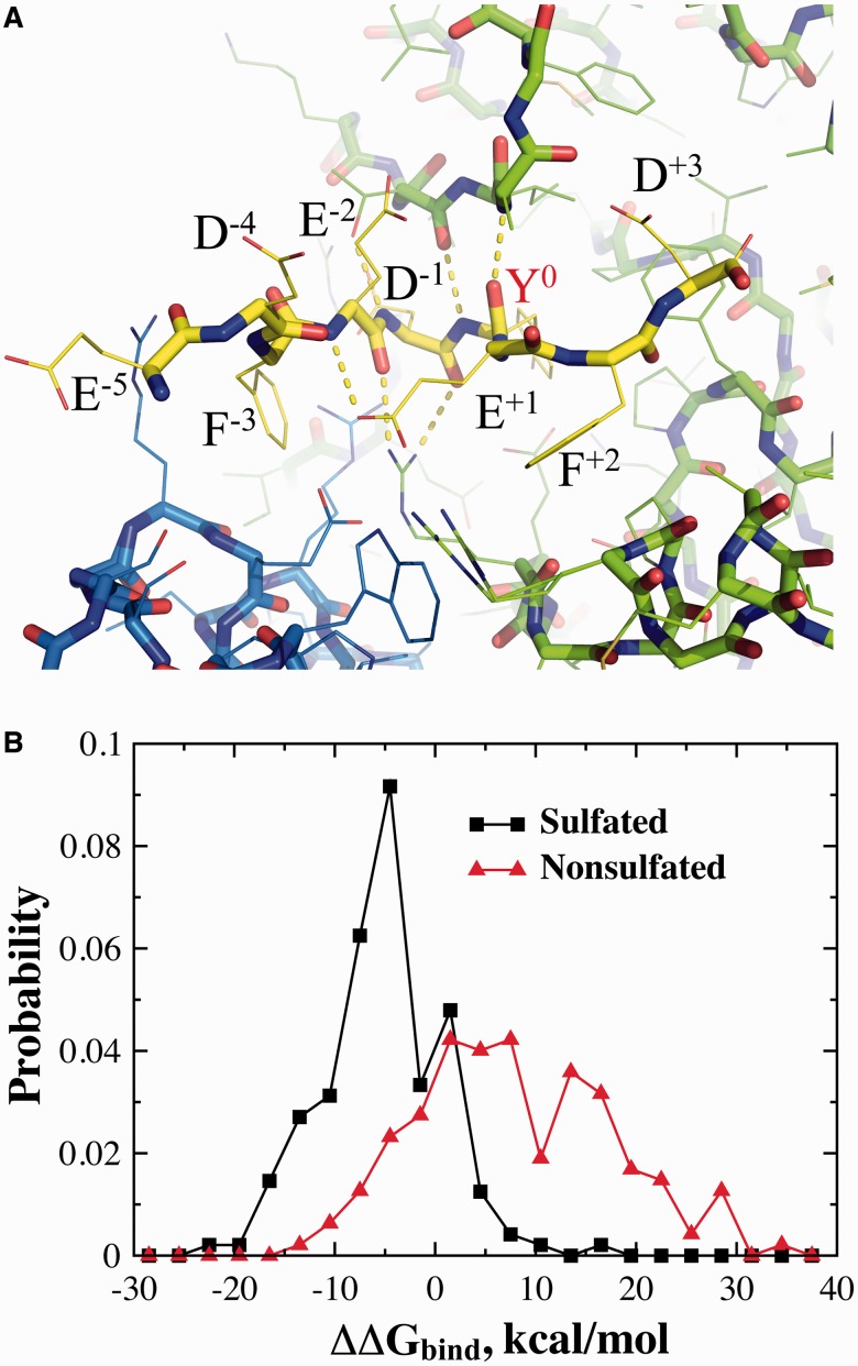
Fig. 2.
The estimation of protein–peptide binding affinity. (A) The structure of the reference nine-residue peptide in the TPST-2 binding pocket as obtained from X-ray crystallography structure, with the sulfated tyrosine positioned as the sixth residue (Y0). The neighboring residues and their positions (superscript) relative to the tyrosine are also marked. (B) The probability density of peptide–enzyme binding scores (ΔΔGbind) for the peptides both sulfated and non-sulfated peptides

