
Figure 3.
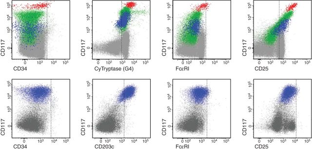
Flow cytometric analysis of mast cell differentiation profiles in the bone marrow of patients with MCL. Bone marrow aspirate samples were obtained from a patient with acute MCL (upper panels; the same case shown in the lower left panel of Figure 2) and chronic MCL (lower panels; case shown in the lower right panel of Figure 2). Expression of cell surface antigens on KIT+ (CD117+) mast cells (events colored blue, green and red) was determined by monoclonal antibodies and multicolor flow cytometry. Mast cells in MCL typically express KIT and lack CD34 (left panels). Mast cells usually also express tryptase and CD203c. However, mast cells in MCL express only low levels of (or no detectable) high-affinity IgE receptors (FcεRI) and only low levels of CD2, whereas CD25 is often expressed on mast cells in MCL.