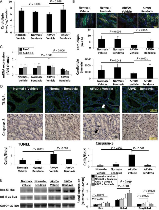
Figure 2.

(A) Stenotic-kidney cardiolipin content decreased in ARVD, but was restored in ARVD + Bendavia (n = 7/group). (B) Fluorescent staining for cardiolipin (red) and cytokeratin (green) confirmed decreased renal expression and staining intensity in ARVD, which was normalized in ARVD + Bendavia (n = 7/group). (C) Expression of Taz-1 mRNA was unaltered, whereas ALCAT-1 was down-regulated in ARVD + Bendavia (n = 7/group). (D) The numbers of TUNEL+ and caspase-3+ cells were elevated in ARVD, yet normalized in ARVD + Bendavia (n = 7/group). (E) Renal protein expression and ratio of Bax and Bcl-xl (n = 6/group).
