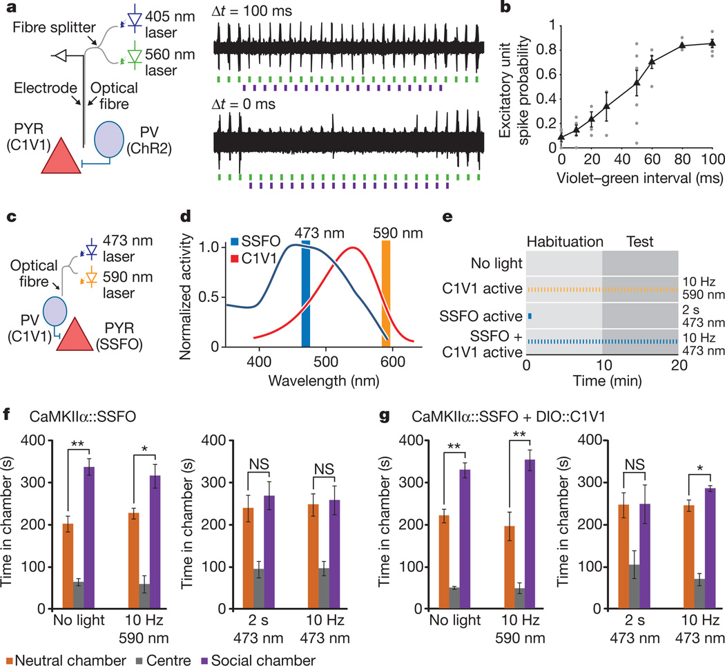
Figure 5. Combinatorial optogenetics enables partial reversal of elevated E/I-balance social behaviour disruption.
a, mPFC optrode recording in an anaesthetized PV::Cre mouse injected with CaMKIIα::C1V1(E162T)-ts–EYFP and Ef1a-DIO::ChR2–EYFP (diagram illustrates experimental setup). Violet (405 nm) light pulses are presented with variable delay (Δt) relative to green light pulses (example traces). b, Summary graph shows probability of green-light-evoked spikes with violet pulses preceding the green light pulses by the indicated delays. Individual points are from single recordings. Black line shows average for all recordings (>3 recording sites per bin). c, Experimental paradigm for SSFO activation in pyramidal neurons and C1V1 activation in PV neurons. d, Action spectra of SSFO (blue) and C1V1(E122T/E162T) (C1V1, red). Orange and blue vertical lines indicate stimulation wavelengths used in the experiments. e, Experiment design and pulse patterns; no-light control was used for baseline behaviour; 2 s 473 nm light for prolonged SSFO activation; 10 Hz 473 nm for co-activation of SSFO and C1V1; 10 Hz 590 nm for C1V1 activation. f, Mice expressing CaMKIIα::SSFO (n = 7) showed significant social preference at baseline, but exhibited social dysfunction after either 2 s 473 nm activation or during 10 Hz 473 nm activation. g, Mice expressing both CaMKIIα::SSFO and DIO-PV::C1V1 (n = 7) showed impaired social behaviour after a 2 s 473 nm pulse, but showed partially restored social behaviour during the 10 Hz 473 nm light stimulation. Activation of C1V1 alone with 10 Hz 590 nm pulses did not impair social behaviour. NS, not significant. Supplementary Fig. 16 shows normal social behaviour under all illumination conditions in YFP-expressing control cohorts. All error bars indicate s.e.m. (*P < 0.05; **P < 0.005).

