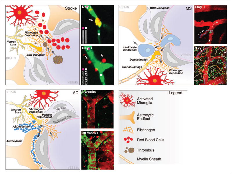
Figure 1.
In vivo imaging of the neurovascular unit in stroke, MS and AD. Schematic representation of the dynamic alterations at the neurovascular unit in different neurologic diseases. Despite different origins, neuropathology and clinical disease progression, there are striking similarities in the cellular processes underlying breakdown of the neurovascular unit. Leakage of the BBB with entry of blood proteins, activation of microglia, astrocyte endfeet retraction, pericyte detachment, neuronal and axonal changes are key elements of cerebrovascular dysfunction. Repetitive in vivo imaging shows extravasation of micro-emboli causing alterations in CBF and restructuring of the microvasculature in stroke,28 migratory paths of T cells after their attachment to the leptomeningeal vessels in a mouse model of MS,45 and gradual progression of cerebral amyloid angiopathy over weeks as captured by time-lapse in vivo imaging in a transgenic mouse model of AD.54 Images reproduced with permission.

