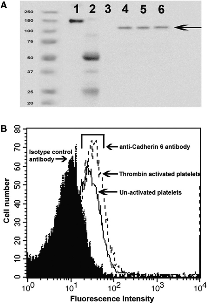
Figure 1.
Cadherin 6 is present on platelets. A, Total platelet lysate from 3 different donors was analyzed by Western blot for the presence of cadherin 6 using a sheep polyclonal antibody against cadherin 6. Lane 1, Cdh6_IgG; lane 2, sheep IgG; lane 3, human IgG; lanes 4–6, total platelet lysate. Cdh6_IgG is composed of the extracellular portion of cadherin 6 fused to the Fc domain of human IgG and therefore has a higher molecular weight than the native protein. B, Platelets were incubated with a polyclonal antibody and analyzed by flow cytometry to confirm the presence of cadherin 6 on the platelet surface. Cdh6_IgG indicates cadherin 6_IgG fusion protein.

