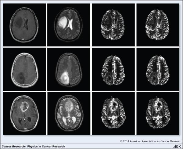
Figure 5.
Grading tumors using DSC-MRI: (L-R) Post Contrast T1, T2, rCBF, rCBV images of a grade II (top row), grade III (middle row) and grade IV (bottom row) tumors generated using DSC-MRI. Note the absence of contrast enhancement in the grade II and grade III tumors. Also, note the elevated rCBV and rCBF in the grade IV tumors

