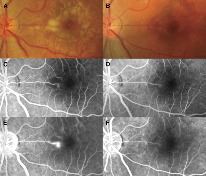
Figure 1.
(A and B) Retinal photographs and (C and D, arterial phase; E and F, late phase) fluorescein angiograms of the left eye. On day 3, increased vessel thickness and tortuosity plus (A) patchy macular whitening with corresponding areas of (C) reduced perfusion and (E) fluorescein leakage were seen. On day 55, normal vessels, (B) no whitening, and (D) normal perfusion around the fovea with (F) no leakage of fluorescein were seen.

