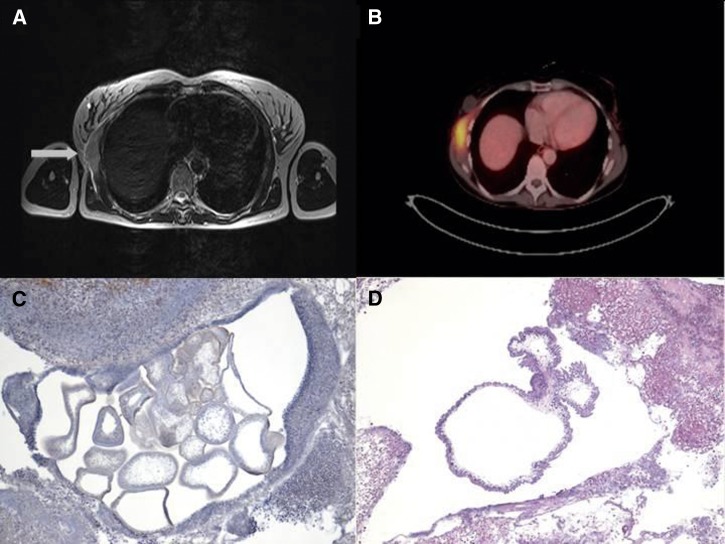
Figure 1.
(A) MRI of the thorax showing the prominent tumor (arrow) at the right upper chest wall located within the chest-wall muscles. T2-weighted image. (B) Positron emission tomography scan showing a pathologic 18F-fluorodeoxyglucose uptake of the tumor. An uptake was also seen in the surrounding lymph nodes (not shown). (C) Histopathological examination of the resected severely inflamed tumor. Multiple sections through several cystic cestode larvae are visible. Original magnification, ×40 (hematoxylin stain). (D) In the center of the inflammatory lesion, a section through the cystic body of a cestode larva is clearly visible. The parasite's tegument appears ruffled and surrounds a spongy stroma. Protruding from the larvae's body, two buds characteristic for T. crassiceps are seen. Differential diagnoses for T. crassiceps cysticercosis include cysticercosis caused by T. solium (single cyst without buds), coenurosis caused by T. multiceps/T. serialis (single cyst with many protoscoleces), and alveolar echinococcosis (multiple cysts but with strong outer laminated layer). Original magnification, ×40 (hematoxylin and eosin stain).

