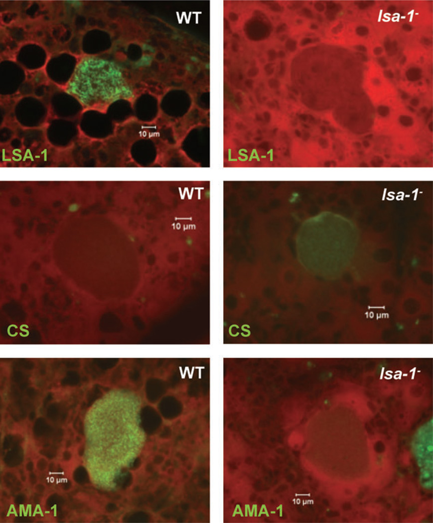
Fig. 5.
lsa-1− late liver stages show protein expression abnormalities. Immunofluorescent micrographs of chimeric SCID/Alb-uPA liver cryosections containing 7-day-old WT or lsa-1− liver stages were stained with parasite-specific antibodies. The micrographs in the top row show that LSA-1-specific antibodies do not bind to lsa-1− liver stages, confirming successful gene deletion. lsa-1− liver stages show abnormal expression of CS protein (middle row micrographs) and AMA-1 protein (bottom row micrographs) when compared with WT liver stages. Scale bar – 10 µm.

