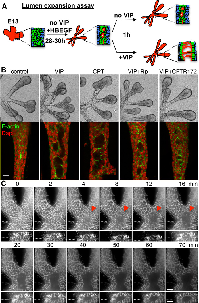
Figure 6. VIP regulates lumen expansion in a cAMP/PKA/CFTR-dependent manner.
(A) Schematic showing lumen expansion assay and observed experimental outcomes. (B) E13.5 epithelia were treated with VIP, cAMP analog CPT-cAMP (CPT; 200 µM), PKA inhibitor Rp-CPT-cAMPS (Rp; 200 µM) or inhibitor of CFTR (CFTR-172, 40 µM) for 1 h. Fluorescent images are single 1µm confocal sections. Scale bar = 10 µm. (C) Epithelia with pre-formed ducts from mice expressing membrane-bound RFP (Tomato) were treated with VIP at time point 0 and lumen expansion was followed for 70 min. Top panels are single confocal XY-sections. The bottom panels are Z-axis reconstructions of 37 individual 1 µm-spaced optical sections. Dashed lines indicate location of optical sections and Z-reconstructions. Arrowhead marks expanding lumen. Scale bars = 20 µm.
See also Figure S5.

