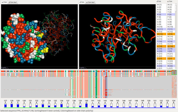
Figure 4.

scTIM/dTIM comparison. The two volume views show the dTIM protein backbone (right), respectively a CPK sphere representation of scTIM (left). The trend image is sorted by the number of common residues. A side-chain polarity coloring is applied, and the two vertical selection paddles are located around position 142. The two residues shown at the bottom (green underline, respectively blue underline) correspond to the two vertical paddle selections.
