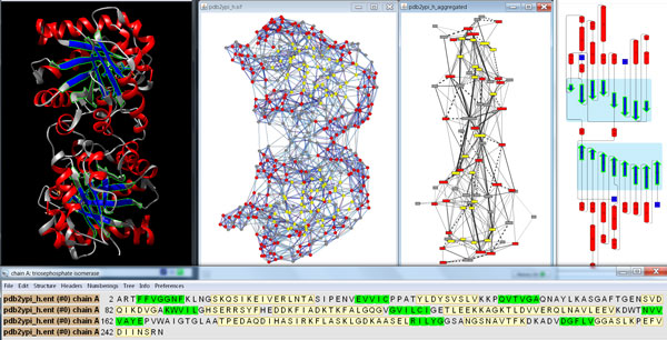
Figure 1.

Simultaneous visualization of biological information using different complementary views. This overview shows the protein structure and the individual residue interactions of the yeast triosephosphate isomerase (scTIM). In particular, the three-dimensional structure and its sequence (top left and bottom, respectively) are shown with UCSF Chimera, the resulting two-dimensional view of the residue interaction network and the aggregated secondary structure network generated with RINalyzer are visualized in Cytoscape (top middle), and the cartoon image of the secondary structure elements is provided by Pro-origami (top right). Residue and network nodes are colored according to their secondary structure (strands in blue and helices in red). Strands that have been selected within UCSF Chimera are indicated by green boundary color in the structure view, by green background in the sequence view, by yellow node color in Cytoscape, and by green boundary color and blue background in Pro-origami.
