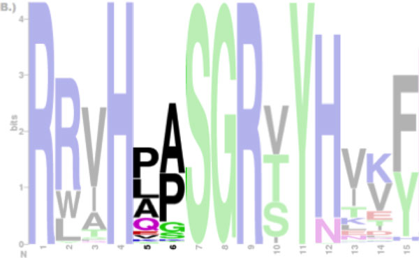
Figure 1.

Sequence logos of the adenylate kinase lid domain from Gram-negative bacteria. It is not clear whether the Proline (P) at position 5 is followed by the Alanine (A) or the Lysine (K) at subsequent position 6. Only positions from 1 through 15 are shown and masked except for position 5 and 6.
