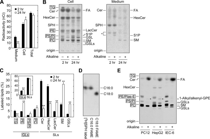
FIGURE 2.
A high proportion of SPH is metabolized to glycerophospholipids. A–C, HeLa cells were labeled with 0.065 μCi of [11,12-3H]SPH for 2 or 24 h. Lipids were extracted from the cells and culture medium. A, the radioactivity associated with the cells and culture medium was quantified using liquid scintillation counter LSC-3600, and data are expressed as the mean ± S.D. from three independent experiments. Statistically significant differences are indicated (t test; *, p < 0.05; **, p < 0.01). B, extracted lipids were treated with or without alkaline solution and separated by normal-phase TLC with 1-butanol/acetic acid/water (3:1:1, v/v), followed by detection with bioimaging analyzer BAS-2500. Cer, ceramide; HexCer, monohexosylceramide; LacCer, lactosylceramide; GSL, glycosphingolipids other than monohexosylceramides and lactosylceramide; SM, sphingomyelin. The glycosphingolipid positioned between S1P and SM and below SM may be globosides Gb3 and Gb4, respectively, considering that these globosides are major glycosphingolipids in HeLa cells (52). The asterisk indicates unidentified lipids. C, the radioactivity associated with each lipid in B was quantified, and data are expressed as the mean ± S.D. relative to the radioactivity in total lipids from three independent experiments. Statistically significant differences are indicated (t test; *, p < 0.05, **, p < 0.01). Inset, radioactivity of total glycerolipids (GLs) and sphingolipids (SLs). D, HeLa cells were labeled with 0.16 μCi of [11,12-3H]SPH for 24 h. Lipids were extracted, treated with alkaline, and subjected to methyl esterification. The generated FAMEs were separated by reverse-phase TLC with chloroform/methanol/water (15:30:2, v/v) and detected by autoradiography. As standards, [9,10-3H]palmitic acid and [1-14C]stearic acid (both from American Radiolabeled Chemicals) were similarly methyl-esterified. E, PC12, HepG2, and IEC-6 cells were labeled with 0.065 μCi of [11,12-3H]SPH for 24 h. Lipids were extracted from the cells, treated with or without alkaline solution, separated by normal-phase TLC with 1-butanol/acetic acid/water (3:1:1, v/v), and detected by autoradiography. Plas-E, plasmanylethanolamine/plasmenylethanolamine; GPE, glycerophosphoethanolamine. Arrowheads represent the breakdown product of Plas-E, 1-alkyl/alkenyl-glycerophosphoethanolamine.

