
FIGURE 4.
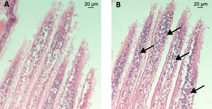
Hemocyte infiltration in gills of LGP32-infected oysters. A and B, histological sections of gills of unchallenged oyster (A) and of oysters infected for 24 h with V. tasmaniensis LGP32 (B). Hematoxylin-eosin staining shows a major hemocyte infiltration in gills of oyster infected with V. tasmaniensis LGP32.