
FIGURE 5.
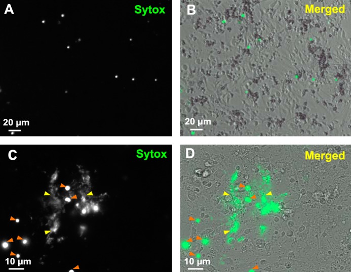
Living oyster hemocytes release extracellular DNA upon bacterial challenge. A–D, cultures of unchallenged hemocytes (A and B) and hemocytes challenged for 45 min with V. tasmaniensis LGP32 (C and D) were stained with Sytox Green and observed by epifluorescence microscopy to reveal extracellular DNA and dead cell nuclei. B and D, bright field images acquired to visualize total cells were merged to epifluorescence images. C and D, extracellular DNA networks (yellow arrowheads) were observed in areas where cells are dead (orange arrowheads) after V. tasmaniensis LGP32 challenge only.