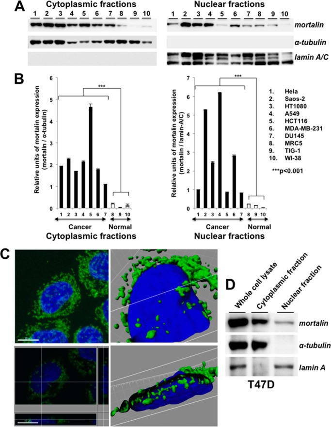
FIGURE 1.
Mortalin is expressed in the nuclei of cancer cells. A, Western blotting of the cytoplasmic and nuclear cell fractions with anti-mortalin antibody showed its presence in the nuclear fractions. Cytoplasmic protein, α-tubulin, and the nuclear protein lamin A/C were used as respective controls for the cytoplasmic and nuclear fractions. B, quantitation of mortalin expression in cytoplasmic and nuclear fractions, S.D. and statistical significance from three independent experiments. C, high-resolution spectral imaging showing mortalin in the cross-section of the nucleus (bottom right panel) of T47D cells. Top left panel, immunostaining of mortalin (green). Nuclei were stained with Hoechst (blue). The white lines in the bottom left, top right, and bottom right panels show the cross-section points. D, Western blot analysis of cytoplasmic and nuclear cell fractions of T47D cells with anti-mortalin antibody showing the presence of mortalin predominantly in the nuclear fraction of cancer cells.

