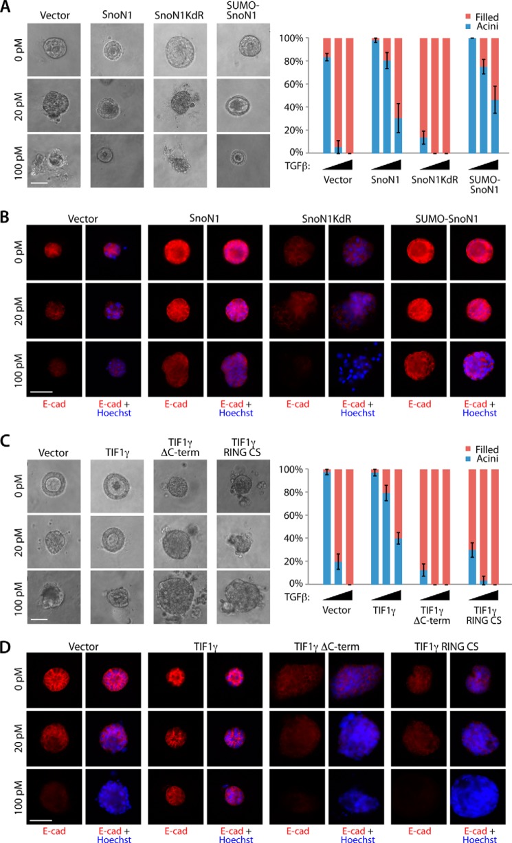
FIGURE 5.
TIF1γ and SnoN1 control epithelial morphogenesis. A, representative DIC images (left panel) and quantification of acini or filled colony morphology (right panel, mean ± S.E., n = 3 or 4) of NMuMG cells transfected with vector control, wild-type SnoN1, SnoN1KdR, or SUMO-SnoN1-expressing plasmids that were left untreated or incubated with 20 pm or 100 pm TGFβ for 10 days. Wild-type SnoN1 and SUMO-SnoN1 significantly suppressed the ability of TGFβ to reduce the proportion of hollow acini (p < 0.05). SnoN1KdR decreased the proportion of hollow acini even in untreated three-dimensional cultures (p < 0.001). B, three-dimensional NMuMG cultures as in A were analyzed as in Fig. 4C. E-cad, E-cadherin. C, representative DIC images (left panel) and quantification of colony morphology (right panel) of NMuMG cells transfected with expression plasmids encoding GFP and wild-type FLAG-TIF1γ, FLAG-TIF1γ RING CS, or FLAG-TIF1γ ΔC-term that were left untreated or incubated with 20 pm or 100 pm TGFβ for 10 days. Wild-type TIF1γ significantly suppressed the ability of TGFβ to reduce the proportion of hollow acini (ANOVA, p < 0.001). Both TIF1γ mutants decreased the proportion of acini with hollow centers even in the absence of TGFβ addition (ANOVA, p < 0.001). D, three-dimensional NMuMG cultures as in C were analyzed as in Fig. 4C. A–C, scale bar = 50 μm.

