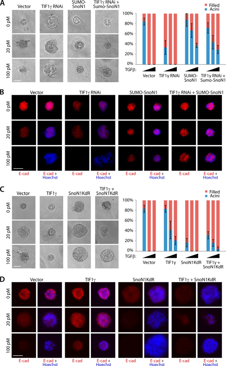
FIGURE 6.
TIF1γ acts via SnoN1 sumoylation to control epithelial morphogenesis. A, representative DIC images (left panel) and quantification of colony morphology (right panel, mean ± S.E., n = 3 or 4) of NMuMG cells transfected with vector control, the RNAi plasmid encoding TIF1γ shRNAs, SUMO-SnoN1 expression plasmid, or the RNAi plasmid encoding TIF1γ shRNAs together with the SUMO-SnoN1 expression plasmid that were left untreated or incubated with 20 pm or 100 pm TGFβ for 10 days. TIF1γ RNAi decreased the proportion of acini with hollow centers even in the absence of TGFβ addition (ANOVA, p < 0.01). SUMO-SnoN1 reversed the ability of TIF1γ RNAi to reduce hollow acini under all three conditions (ANOVA, p < 0.05). B, three-dimensional NMuMG cultures as in A were analyzed as in Fig. 4C. E-cad, E-cadherin. C, representative DIC images (left panel) and quantification of colony morphology (right panel) of NMuMG cells transfected with the vector control, expression plasmid encoding wild type TIF1γ, SnoN1KdR, or TIF1γ together with SnoN1KdR that were left untreated or incubated with 20 pm or 100 pm TGFβ for 10 days. SnoN1KdR suppressed the ability of wild-type TIF1γ to maintain the proportion of hollow acini in the absence and presence of 20 pm TGFβ (ANOVA, p < 0.05). D, three-dimensional NMuMG cultures as in C were analyzed as in Fig. 4C. A–C, scale bar = 50 μm.

