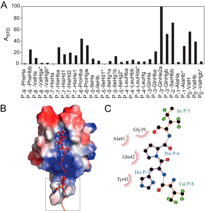
FIGURE 5.
Binding of PKCα to PICK1. A, normalized STD amplification factor (ASTD) measured for the 1H resonances in the PKCα C12 peptide. B, HADDOCK docking model of the PKCα C12 peptide based on the amplification factors in the STD experiment. In the single cluster of structures obtained from the calculation, the C terminus of PKCα binds PICK1 in the canonical binding mode, which is further stabilized by an upstream binding site involving residues located in the Cys-44–Pro-45–Cys-46 βB-βC loop and ligand residues His P−7 and Pro P−6. The PDZ domain is colored with the electrostatic potential from −2 kT/e (blue) to 2 kT/e (red). C, highlight of upstream binding contacts.

