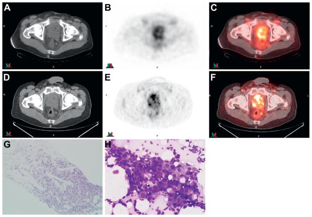
Figure 3.
Imaging in 61-year-old patient after external beam radiation therapy and hormonal therapy with increasing PSA to 1.96 ng/ml reveals extensive biopsy proven recurrent disease in prostate and multiple pelvic nodes. 111In-capromab pendetide CT (A), scintigraphy (B) and fused image (C ) show abnormal uptake in prostate and left perirectal node. Anti-3-[18F]FACBC CT (D), PET (E ) and fused image (F ) at same level also show abnormal uptake in prostate and left perirectal node. Prostate core biopsy demonstrates prostatic Gleason 4 + 4 = 8 adenocarcinoma (G). H&E, reduced from × 10. Fine needle aspiration of perirectal node demonstrates malignant prostate adenocarcinoma cells with glandular formation and prominent nucleoli (H ). 111In-capromab pendetide findings were considered abnormal in node but there was better lesion contrast on anti-3-[18F]FACBC imaging with more nodes identified in pelvis. Diff-Quik stain, reduced from ×40.

