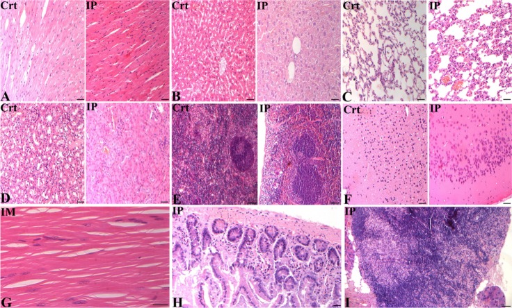
Figure 5.
Photomicrographs of different organs collected from euthanized nude mice, four weeks post amnion-derived mesenchymal stem cell injection via the intramuscular (IM) and intraperitoneal (IP) routes. (A) Longitudinal section of cardiac muscle showing a common arrangement of muscle fibers with centralized nuclei. (B) Photomicrograph of the liver, central vein and numerous hepatic cords lining the sinusoidal capillaries. (C) Lung morphology, pulmonary alveoli, and alveolar ducts. (D) Cross-section of the kidney and overview of the glomeruli and renal tubules. (E) Cross-section of the spleen, normal distribution of lymphatic nodules, trabeculae, and white and red pulp. (F) Brain photomicrograph showing different layers of the cerebral cortex. (G) Longitudinal section of thigh skeletal muscle, nuclei in periphery of muscle fibers, and numerous transverse striations. (H) Small intestine, numerous glands immersed in the submucosal tissue. (I) Normal distribution of medullary cords and medullary sinuses of lymph node, with adipocytes in the periphery. (A, B, C, E and G) Bars 20 μm (40×), (D, F and H) 20 μm (20×), and (I) 50 μm (20×).
Abbreviation: Ctr, control.

