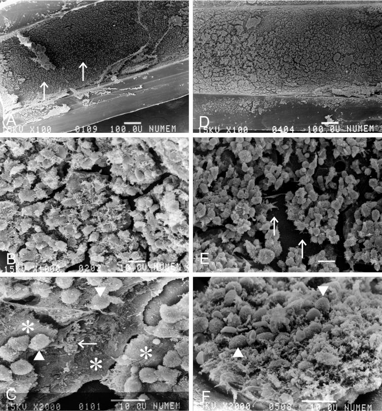
FIG. 3.
Scanning electron microscopy of the biofilm on the inner space of the inoculation tube (longitudinal section) in mice that received 40 days of saline treatment after inoculation with P. aeruginosa (A, B, and C) and 40 days of ERY treatment (D, E, and F). Bars, 100 μm (A and D) and 10 μm (B, C, E, and F). The arrows in panel A show the smooth surface of the biofilm covering the inner wall of the tube. The inflammatory cells (arrowheads) are separated from the bacteria (arrow) embedded deeply in the multilayer biofilm (asterisks) (C). The arrows in panel E show the surface of the base layer of the biofilm. In mice treated with ERY, the inflammatory cells (arrowheads) were mixed with the biofilm (F).

