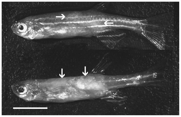
Figure 1.

Zebrafish infected with P. hyphessobryconis. Upper image shows mottled appearance with light areas (arrows) on flanks. Lower image is same fish with skin removed, exhibiting opaque regions in muscle (arrows) representing massive infection. Bar = 500 μm.
