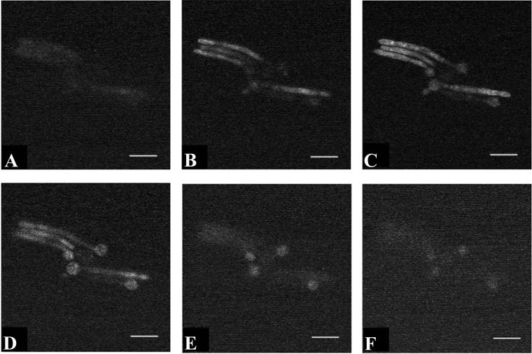
FIG. 2.
Cellular localization of Photofrin in C. albicans. Photomicrographs obtained by fluorescence confocal microscopy are depicted from a series of 1-μm-thick optical sections through viable C. albicans strain 3153A cells after uptake of Photofrin. Panels A to F were obtained in sequence through the same microscopic field. Fluorescence was visualized throughout the cell, suggesting that the compound had reached the cell interior. Bar, 15 μm.

