FIGURE 3.

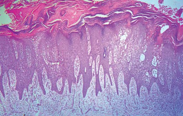
Acanthotic epidermis with regular elongation of interpapillary cones and atrophy in sections covering the dermal papillae. Focal spongiosis with intraepidermal vesicles. Hyperorthokeratotic stratum corneum, with Munro’s microabscesses

Acanthotic epidermis with regular elongation of interpapillary cones and atrophy in sections covering the dermal papillae. Focal spongiosis with intraepidermal vesicles. Hyperorthokeratotic stratum corneum, with Munro’s microabscesses