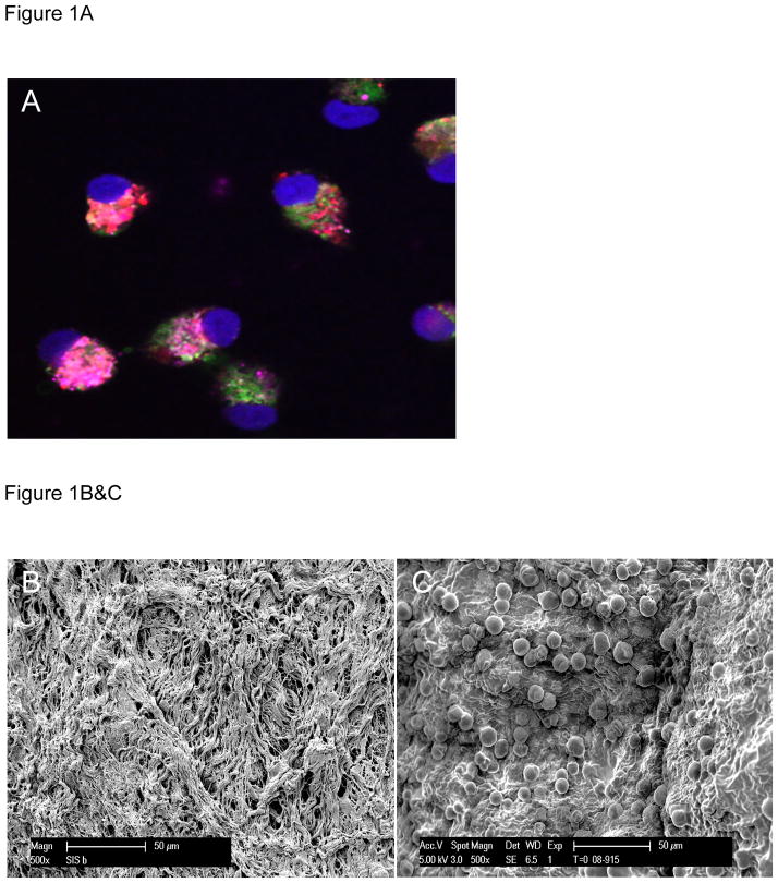
Figure 1.
Figure 1A- At day seven in culture, isolated MNCs grown on extracellular matrix show universal expression of VEGFR2 (pink), stain positive for Isolectin (green), and show uptake of Di-Iodinated acetyl-LDL (red), all of which are characteristic of EPCs (nuclei are stained with DAPI-blue).
Figure 1B&C- Scanning electron micrographs of ECM before (B) and after (C) seeding with EPCs show densely populated, adherent cells

