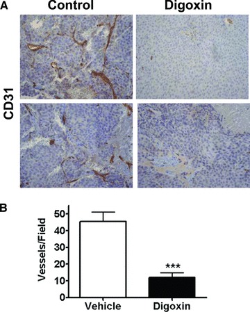
Figure 3.

Digoxin inhibition of blood vessel density in C4‐2 xenogaft tumor. Nude mice were injected subcutaneously with 1× 10⁶ C4‐2 cells in a 1: 1 ratio with matrigel. Tumors were established and upon reaching 7mm in diameter were castrated. Mice were allowed to recover for 10 days before starting intraperitoneal treatment with either digoxin (2mg/kg) or vehicle alone for 7 days. Tumors were harvested 2 days after treatment stopped. IHC was performed on the tumor tissue for CD31 to view microvascular density. (A) Displays a representative view at 20×. (B) Shows the scoring of the tumor tissue counting CD31 positive vessels in each field. The graph displays the average of six random fields per mouse with four mice in each group. The digoxin treated group had a significant fourfold reduction in the number of blood vessels (***p= 0.0004).
