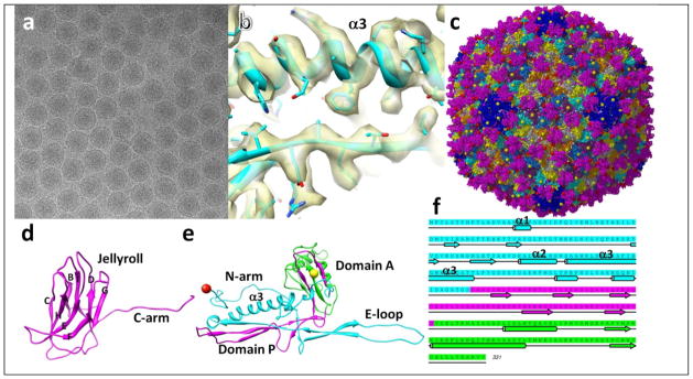
Fig. 1. Structure of BPP-1 at 3.4Å resolution determined by cryoEM.

(a) cryoEM image of BPP. (b) Density of a portion of MCP protein. (c) atomic model of BPP capsid. Balls indicate Thr6 (red) and Val330 (yellow) exposed on the external surface of the BPP capsid. (d) ribbon model of the cement protein. (e) ribbon model of MCP. The three building blocks were indicated by cyan (N-terminal), purple (two strands in domain P, and two strands in domain A), and green (major part of domain A). (f) amino acid sequence and secondary structure of MCP, with segments colored according to structures of the building blocks in (e).
