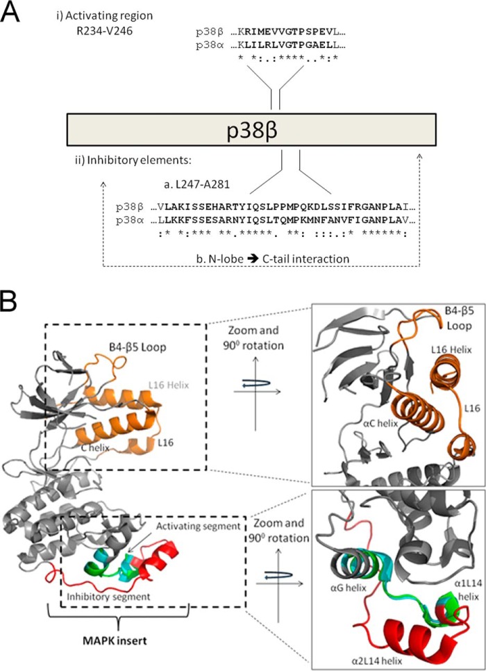
FIGURE 6.
A schematic (A) and structural (B) representation of regions that regulate p38β autophosphorylation. The crystal structures of p38α (Protein Data Bank code 4E5B; gray backbone) and p38β (Protein Data Bank code 3GC8; backbone not shown) were aligned according to the Arg234–Val246 region, p38β numeration, and are presented from two angles rotated 90°. (i) A and B, the unique intrinsic autophosphorylation activity of p38β is triggered by the Arg234–Val246 region composing part of the α-G helix and MKI. B, the G-MKI region of p38α is shown in cyan, and that of p38β is shown in green. (ii) A and B, p38β intrinsic autophosphorylation is suppressed in mammalian cells via part of the MKI (shown in red in B) and depends on an interaction between the N-lobe and C-tail. The C-tail, composed of L16 and the L16 helix, the N-lobe elements that interact with the C-tail, the C helix, and the β4-β5 loop, is shown in orange.

