FIGURE 9.
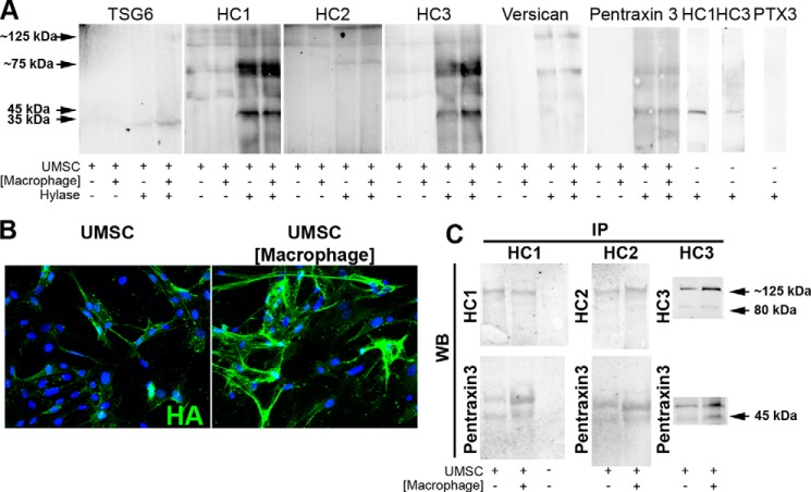
UMSCs express a rich HA/TSG6/HC/pentraxin 3 ECM. UMSCs were placed in co-culture with inflammatory cells seeded in a transwell insert with a 0.44-μm pore. Proteins were extracted and digested or not with Hylase prior to analysis by Western blotting. A, TSG6, HC1, HC2, HC3, versican, and pentraxin 3 were only detected when the protein lysate was digested with Hylase. B, HA staining (green) in UMSCs and UMSCs exposed to inflammatory cells seeded in transwell inserts. C, immunoprecipitation was done with the conditioned medium from UMSCs and UMSCs exposed to inflammatory cells with anti-HC1 and anti-HC2 and subjected to Western blotting with anti-HC1, anti-HC2, and anti-pentraxin 3. The nuclei were stained with DAPI.

