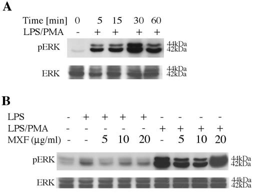
FIG. 2.
Activation of ERK1/2 in LPS- and LPS-PMA-stimulated THP-1 cells and effects of MXF. (A) Time-dependent studies. THP-1 cells were incubated in serum-free medium and exposed to 1 μg of 100 nM LPS-PMA per ml for the indicated times. (B) Effects of MXF on LPS- and LPS-PMA-induced activation of ERK1/2. THP-1 cells were preincubated in serum-free medium in the absence or the presence of the indicated concentrations of MXF and then treated with LPS or LPS-PMA for 30 min. The cytoplasmic extracts were prepared and subjected to Western immunoblotting with an antibody specific for phosphorylated form of ERK1/2 (upper blots in panels A and B) and total ERK1/2 (lower blots in panels A and B). The experiment was repeated three times, and each experiment produced similar results.

