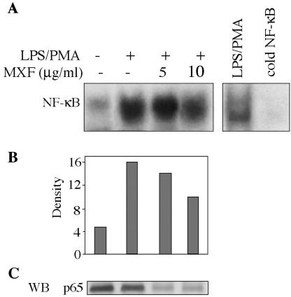
FIG. 6.
Effects of MXF on LPS-PMA-induced activation of NF-κB. (A) THP-1 cells were preincubated in serum-free medium in the absence or presence of MXF and then treated with LPS-PMA for 2 h. Nuclear extracts were prepared and assayed for NF-κB as described in the legend to Fig. 5. (B) Bands were quantified by optical densitometry. (C) Expression of p65 NF-κB protein in LPS-PMA-stimulated cells. The Western blot (WB) illustrates the expression of the p65 NF-κB protein in nuclear extracts from cells exposed to LPS-PMA and preincubated in the absence or presence of MXF. Representatives of the blots from two experiments are shown.

