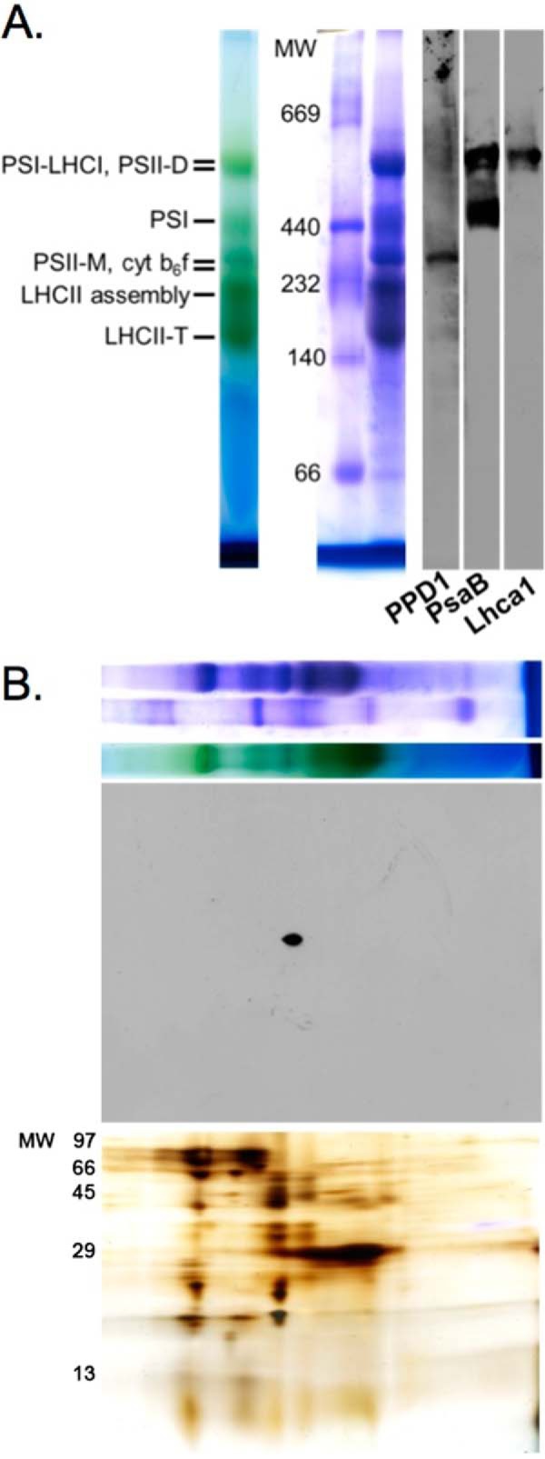
FIGURE 5.

Detection of PPD1 in a protein complex. A, BN-PAGE separation of dodecyl β-d-maltoside-solubilized wild-type thylakoid membranes (unstained, Coomassie-stained, and immunodetection of PPD1, PsaB, and LhcaI proteins). B, two-dimensional (BN-LiDS) PAGE separation of wild-type thylakoids with the first dimension BN-PAGE panel (top) shown above the PPD1 immunodetection panel (middle) and silver-stained panel (bottom). cyt, cytochrome; M, monomer; D, dimer; T, trimer.
