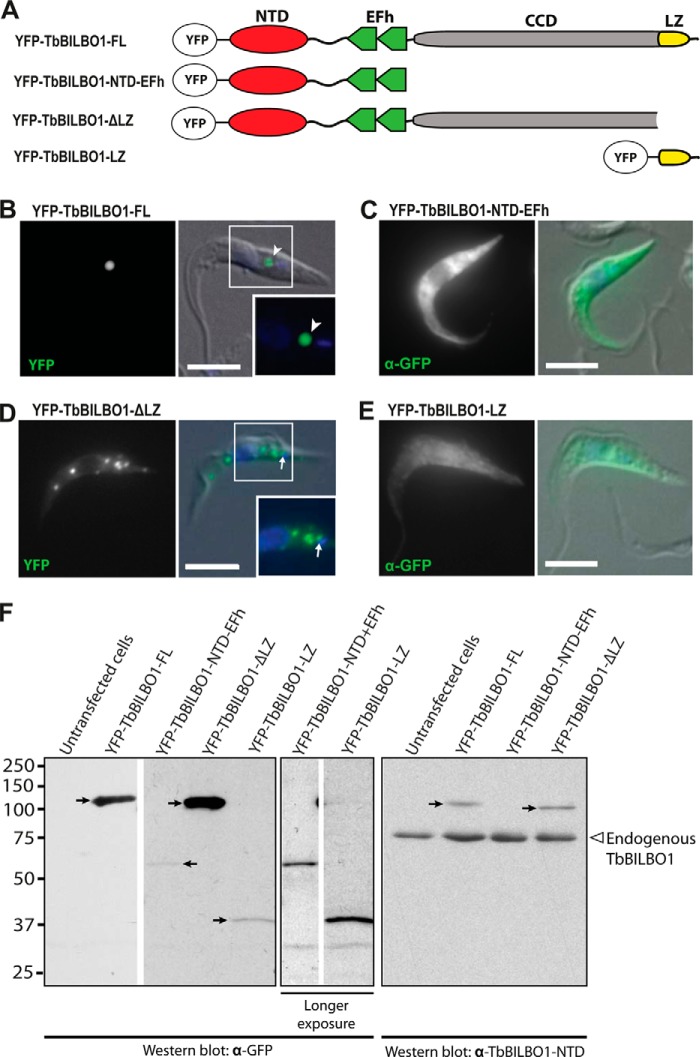
FIGURE 2.
The TbBILBO1-LZ is necessary but not sufficient for targeting to the FPC. A, TbBILBO1 truncation constructs used for the experiment. B–E, localization of YFP-tagged TbBILBO1 truncations in transiently transfected T. brucei cells. Fluorescence microscopy was used to visualize protein localizations. For YFP-TbBILBO1-NTD-EFh and -LZ, low expression levels precluded observation of YFP directly, and anti-GFP antibodies were used to visualize the protein instead. DAPI (blue) was used to stain DNA. The insets in B and D, indicated by white-outlined boxes, are enlargements of the smaller boxed areas. Scale bars: 5 μm. F, immunoblots of whole-cell lysates from transiently transfected T. brucei cells probed using anti-GFP antibodies or anti-TbBILBO1-NTD. The YFP-tagged proteins are indicated by arrows. Longer exposures were used for YFP-TbBILBO1-NTD-EFh and -LZ to better visualize the target proteins. Endogenous TbBILBO1 proteins are marked by an empty arrowhead. The YFP-TbBILBO1-LZ construct is not detectable with the anti-TbIBLBO1-NTD antibodies, as it does not contain the NTD.

