
FIGURE 8.
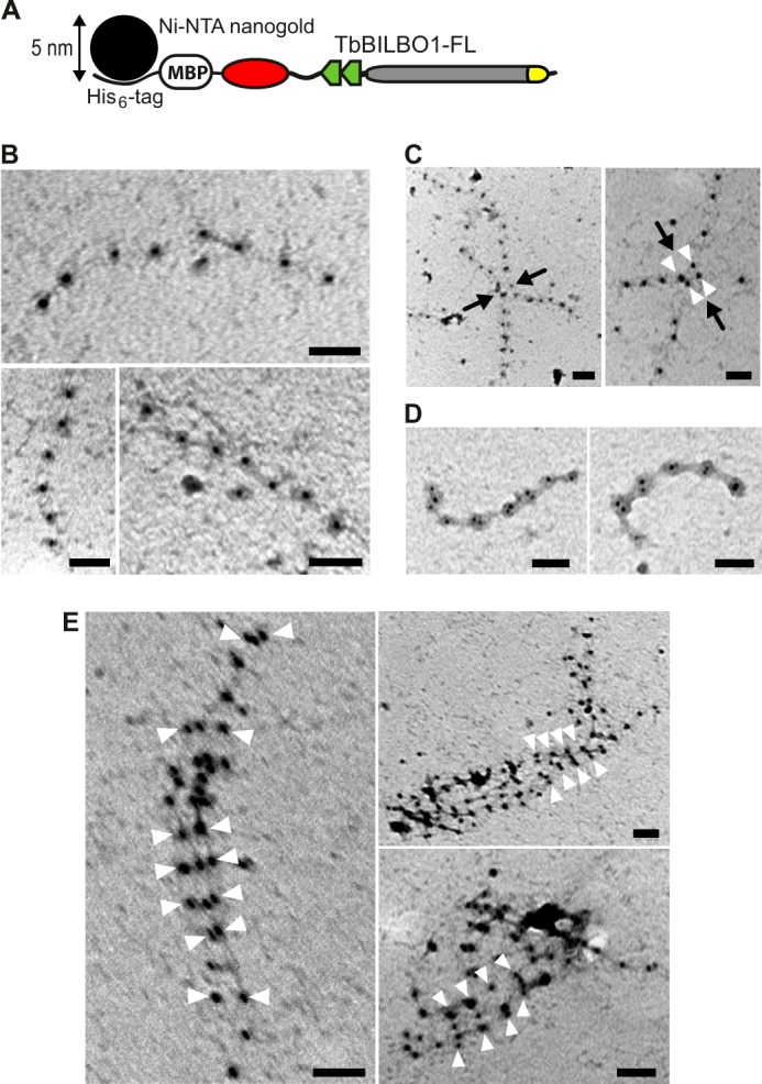
Linear and lateral assemblies of full-length TbBILBO1 revealed by Nanogold labeling-coupled negative staining EM. A, schematic showing the binding of the Ni-NTA-Nanogold particle to the His6 tag at the N terminus of MBP in the fusion protein. B, negative staining EM image of His6-MBP-TbBILBO1-FL labeled with 5-nm Ni-NTA-Nanogold. The oligomers are single linear filaments. C, lateral interactions of two filaments. Black arrows indicate the junctions of the interactions. White arrowheads mark the paired Nanogold particles. D, laterally associated two-filament structures. E, lateral assembly of multiple filaments. White arrowheads mark the clustered Nanogold particles in register. Scale bars: 50 nm.