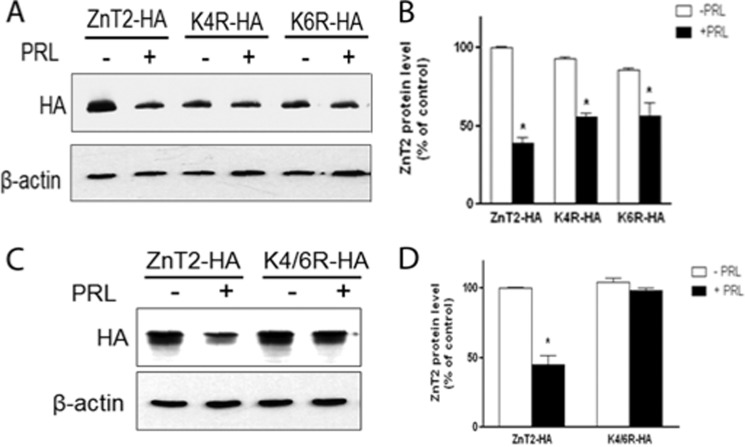
FIGURE 5.
PRL-stimulated ZnT2 degradation requires two lysine residues in the N terminus. A, representative immunoblot of ZnT2-HA in cells expressing ZnT2-HA, Lys4-HA, or Lys6-HA treated with PRL (+) compared with control (−) cells. Transfected cells were pretreated with cycloheximide and then treated with or without PRL for an 8 h, and the levels of each mutant protein were detected by anti-HA antibody. C, representative immunoblot of ZnT2-HA in cells expressing ZnT2-HA or K4R/K6R-HA treated with PRL (+) compared with control (−) cells. Transfected cells were pretreated with cychloheximide and then treated with or without PRL for 8 h, and the levels of each mutant protein were detected with anti-HA antibody. B and D, band intensity of ZnT2-HA normalized to β-actin expressed as a percentage of untreated cells. Data represent the mean percentage of ZnT2 protein level relative to untreated cells expressing ZnT2-HA ± S.D. (error bars) from two independent experiments. *, significant effect of PRL treatment on ZnT2 protein level, p < 0.05.

