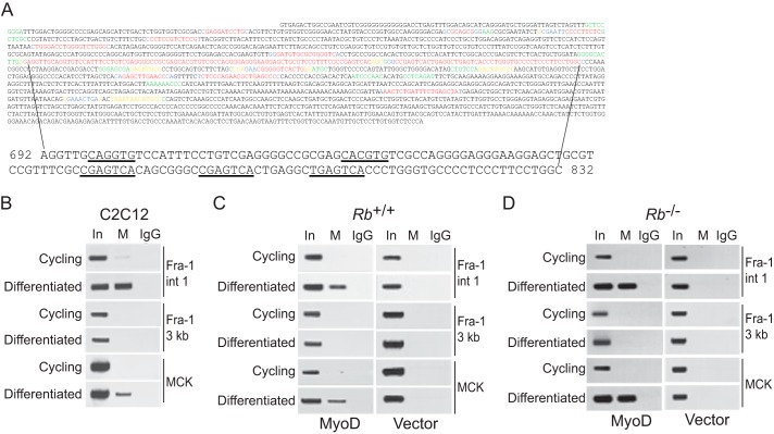
FIGURE 4.
MyoD is localized to E boxes located in the intronic enhancer of Fra-1 during differentiation. A, first 2000 bp of murine and human intron 1 of Fra-1 were analyzed with Bayes block aligner (top). Probability of conservation: 0.8–0.999 (red), 0.6–0.799 (blue), 0.4–0.599 (yellow), 0.2–0.399 (green), and 0–0.199 (black). The largest region showing the highest degree of conservation is shown at the bottom, and E boxes and AP-1 sites are underlined (coordinates are defined by setting the first nucleotide of intron 1 to 1). B, MyoD localization to intronic E boxes of Fra-1 during differentiation. ChIP analysis of cycling and differentiated C2C12 cells was performed with antibody to MyoD (M) or IgG as control. For the precipitated DNA, sequences spanning the E boxes in intron 1 of Fra-1 (Fra-1 int 1), ∼3 kb downstream of the Fra-1 intronic E boxes (Fra-1 3 kb), and the MCK enhancer (MCK) were analyzed by PCR. One-tenth of the lysates used for ChIP were also subjected to PCR (input, In). The results are representative of at least three independent experiments. C, as in B, except Rb-positive fibroblasts were infected with MyoD-encoding virus (left panel) or empty vector (right panel). The results are representative of at least three independent experiments. D, as in C, except Rb-deficient fibroblasts were employed. The results are representative of at least three independent experiments.

